Sample Projects
I'm currently enrolled in an interdisciplinary Doctoral Training Program in Biomedical Imaging. During our first year, we complete a set of short courses to quickly develop skills in various areas. Alongside our lectures, some of the courses use short 'hands-on' projects to help us learn by 'doing'. Below are some examples of the projects that I've been involved with. I'll update this periodically.
Construction of a Basic Radio Frequency Coil
We built a radio frequency receiver coil to acquire an image of a spherical phantom. February 1. Left: The resulting phantom images in the coronal, sagittal, and transverse plane from a 3D gradient echo sequence on the Phillips 7T. Right: The finished coil situated on top of the spherical phantom consisting of H2O, CuSO4 and NaCl.
Team: C Hill, E Bluemke
Microscopy Auto-focus Project
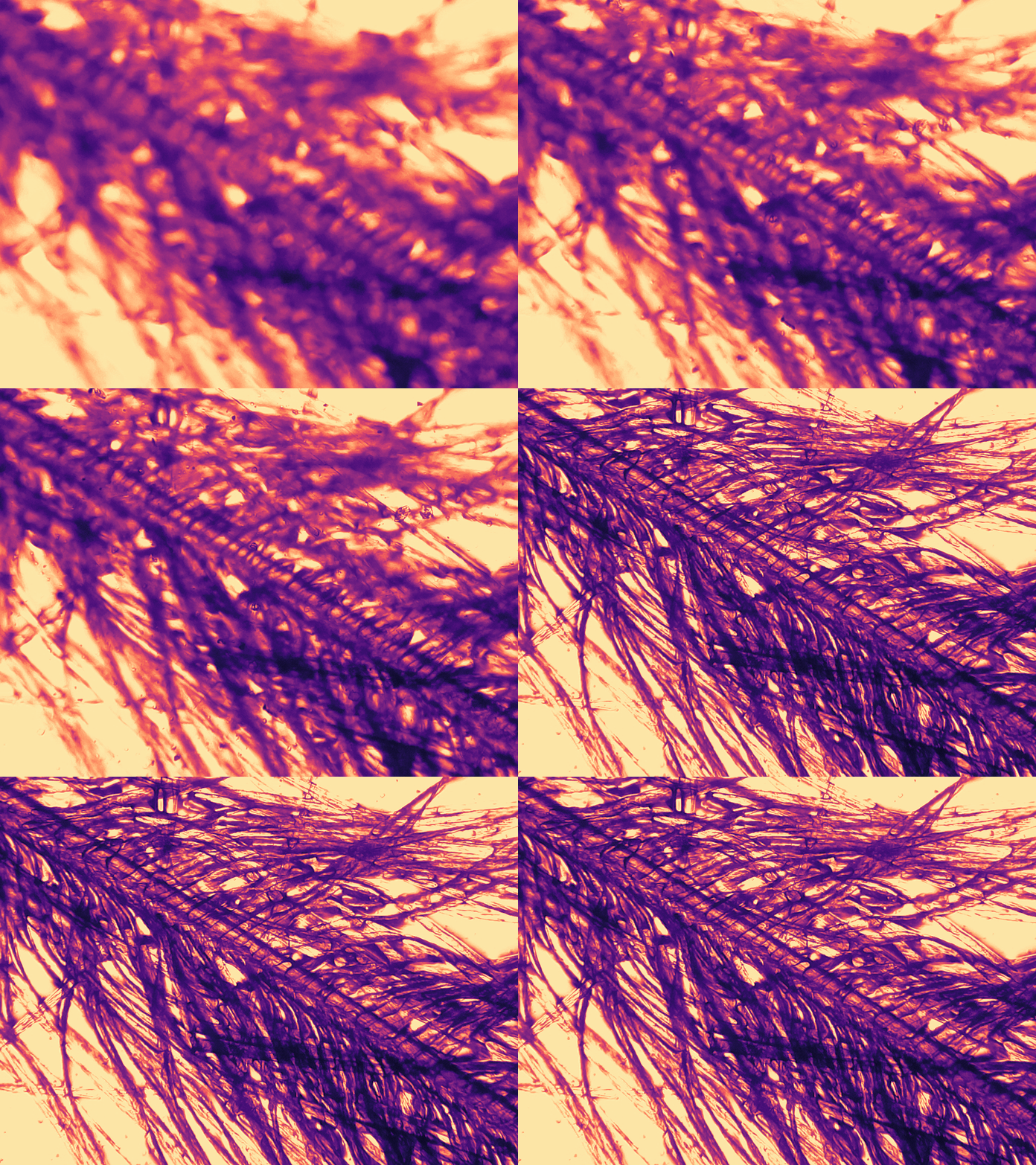
Automated focusing of a simple upright microscope using a Raspberry Pi. January 16-19. Task: design and implement an autofocus algorithm in Python and use this to drive the stepper motors in a feedback loop to acquire optimal focus in bright field and fluorescent microscopy.
Team: G Belsley, C Millard, E Bluemke
Variational Bayes in Neural Networks
Implementation of a variational autoencoder to reproduce the results presented by Kingma and Welling in Python using MNIST as a test dataset. January 2-12. In the report, we demonstrate variational inference implemented within an autoencoder. An autoencoder is a type of neural network in which the input is encoded into latent space through an information bottleneck and subsequently decoded to reconstruct the input. Variational inference methods are used when the posterior distribution cannot be evaluated, so an approximate distribution is chosen from which to sample from, optimizing the parameters of the approximate distribution to best fit the true posterior.
Team: C Wild, R Stephens, E Bluemke
Automated Glioma Segmentation in Multimodal MRI
Support Vector Machine Classification for Segmentation of Gliomas in Pre-Operative Multimodal MRI. December 11-14. Due to their highly heterogeneous appearance, segmentation of gliomas in multimodal MRI scans remains a challenging task in medical image analysis. This automatic brain tumour segmentation method uses a support vector machine classifier to segment tumour tissue from health brain tissue based on multimodal voxel intensity.
E Bluemke
Automated Coin Counter
Automated coin identification and value calculation using a Raspberry Pi. November 6-10. In this project, we developed a fairly reliable script that would use a Raspberry Pi to automatically capture an image, identify all coins in the image, and calculate and display the value of the coins to the user. Works on both GBP and CAD.
Team: G Hutchinson, E Bluemke


